Can Synthetic Melanin Protect Our Skin? My Research Journey at IIT Bombay
By LifETIME CDT Student: Mohamed Touhid Patel (University of Birmingham)
Where my placement took place?
This summer, I had the opportunity to join M-LAB (Matrix, Materials, Mechanics, and Microfluidics Lab- https://iitbombaymlab.wixsite.com/mlab) at the Indian Institute of Technology Bombay, Mumbai, within the Department of Chemical Engineering. I worked under the guidance of Prof. Abhijit Majumder (https://x.com/abhijit_MLab?ref_src=twsrc%5Egoogle%7Ctwcamp%5Eserp%7Ctwgr%5Eauthor), whose pioneering research spans organ-on-chip models, microfluidics, and gradient devices for studying cancer, alongside a deep commitment to scientific outreach. His group explores how fundamental science—like cell mechanics and microfluidic systems—can answer pressing biomedical questions.
What the project aimed to achieve?
During my placement, we designed a project titled: Investigating the Role of Synthetic Melanin in Modulating Oxidative Stress: From 2D Skin Models to Human Skin Validation.
The aims were threefold:
- To study the uptake of synthetic melanin by human keratinocytes (HaCaT cells).
- To investigate how synthetic melanin modulates oxidative stress and DNA damage.
- To explore its effect on wound healing, adhesion, and barrier function in in-vitro models (2D and 2.5D).
In the long term, this work will be validated at the University of Birmingham, with Prof. Pola Goldberg Oppenheimer (ANMSA Research Group), expanding the findings into human skin biopsies.
How I carried out the project?
The first step was training in cell culturing, which gave me the foundation to work with HaCaT keratinocyte cells—the central model for our study.
From there, my responsibilities included designing and carrying out experiments such as:
- Oxidative stress assay (DCFDA): To see how melanin influences reactive oxygen species (ROS) scavenging.
- DNA damage assessment: Using γ-H2AX staining to detect double-strand breaks caused by oxidative stress.
- Adherence junction analysis: Via E-Cadherin staining to examine how melanin affects cell-cell adhesion.
- Wound healing assay: The classic “scratch assay” to track how quickly keratinocytes close a wound gap with and without melanin.
- Barrier integrity study: Using the FITC-D model to evaluate how melanin impacts skin barrier function.
Every experiment offered both challenges and insights.
Why it matters?
Melanin isn’t just the pigment that gives our skin its colour—it also plays a protective role, especially in neutralizing oxidative stress. Excess oxidative stress contributes to DNA damage, premature aging, and skin disorders.
By studying synthetic melanin, our project aims to answer: Can externally supplied melanin support skin health, especially in patients with conditions where melanin is lacking or dysfunctional?
If successful, this approach could open new doors for managing conditions like psoriasis, actinic keratosis, and vitiligo, where oxidative stress and impaired barrier function play a central role.
Ultimately, the hope is that synthetic melanin could serve as a therapeutic antioxidant shield, scavenging harmful ROS, protecting DNA, and supporting wound healing—bringing science from the lab bench one step closer to helping patients.
Looking ahead
My placement at IIT Bombay was more than just lab work—it was a glimpse into how basic cellular experiments can evolve into clinical insights. With the next phase of validation at the University of Birmingham, this research could contribute to a new class of skin therapies.
For me personally, it was also a lesson in patience, precision, and the excitement of watching cells under the microscope—knowing that every tiny change could lead to a bigger story for human health.
Image 1.
A. Photograph taken at the Central Library, IIT Bombay.
B. Group Photograph taken at the M-LAB internal facility, IIT Bombay.
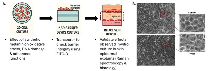
Image 2.
A. Schematic illustrating the workflow of the study, moving from 2D skin models to human skin explants, and highlighting the role of synthetic melanin in modulating oxidative stress. B. Phase-contrast microscopy image showing the internalization of synthetic melanin in HaCaT cells.