$1 Billion Concussion
By LifETIME CDT Student: Matthias Lim (University of Birmingham)
Repetitive head injury has been shown to result in cognitive impairment of many athletes, including early onset dementia, Parkinson’s Disease, and Alzheimer’s Disease. Sporting organisations, therefore, have a critically important ethical duty to actively monitor the health and recovery of their athletes, monitor, and to rapidly diagnose when something has gone wrong preferably, at the point-of-injury. The NFL has awarded nearly $1Bn in compensation as part of a settlement program for failing to warn and protect its players from repetitive head injuries.
My project is focused on developing and optimising a novel approach to diagnosing and monitoring traumatic brain injury (TBI) at the point-of-care. A single drop of blood from a finger prick is all that is required to identify whether a patient has a TBI.
To that end, two key components are required:
- A microfluidic device to separate blood cells from blood serum
- A spectroscopic Raman probe and a Smart substrate.
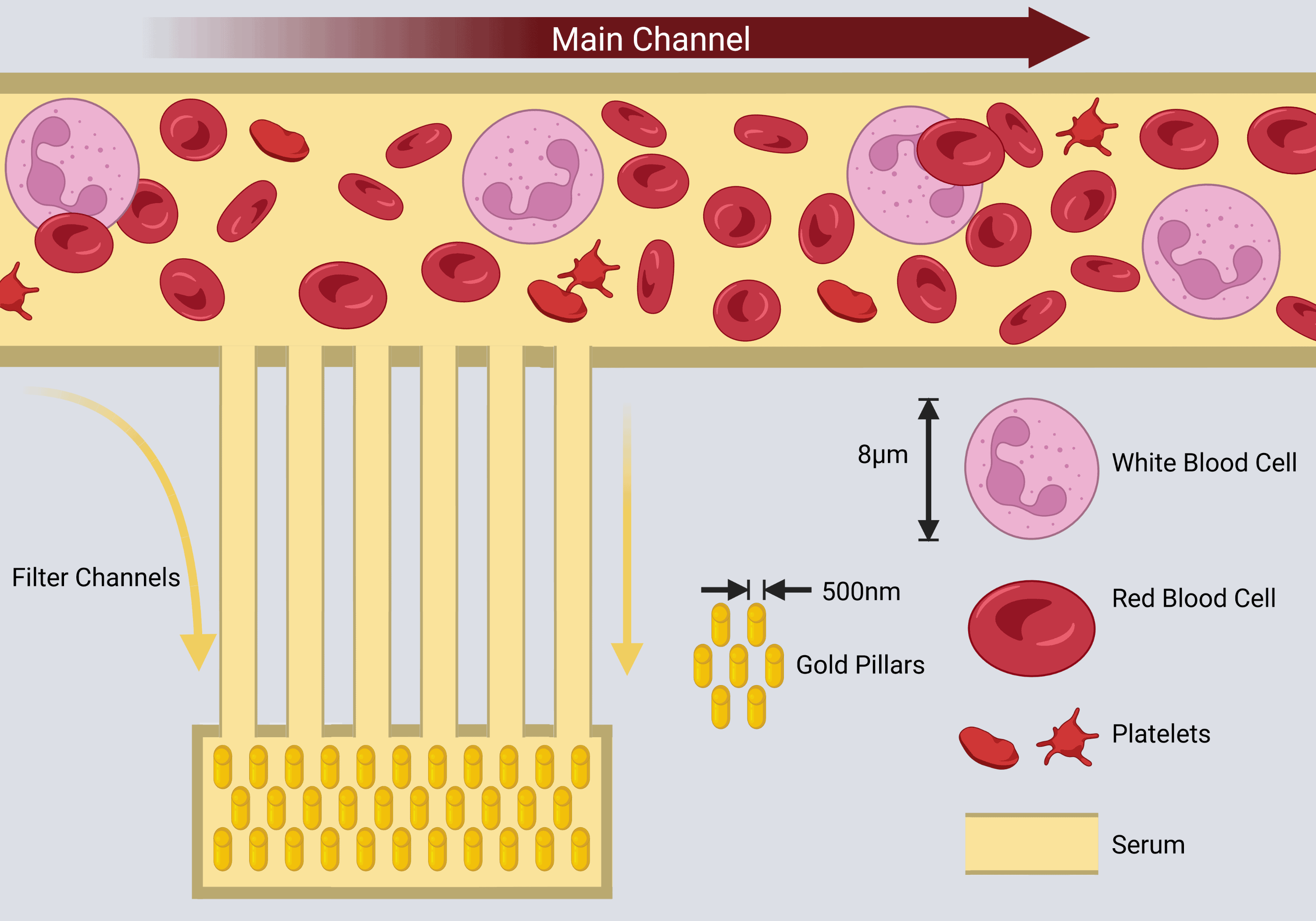
Figure 1: Schematic of the “combs” microchannel design (Rickard et al., 2020). A drop of blood is placed into the inlet reservoir, and capillary forces draw the whole blood through the channels. Small filter channels filter out red blood cells by size separation. Scales are approximate. Created with BioRender.com
As illustrated in Figure 1, a sample of whole blood is deposited into the lab-on-a-chip device. Capillary forces draw blood along the main channel. Small filter channels (“combs”) do not allow large biological material such as red blood cells, white blood cells, and platelets to pass through, however, blood serum is able to flow through. These small channels then deliver filtered serum to the smart Raman substrate.
The primary prototyping technique used in my research is UV lithography. This technique allows me to rapidly produce structures that are smaller than 1% of the thickness of a human hair. An example of these structures is shown in Figure 2, where pillars approximately 500nm in diameter were produced using this method.
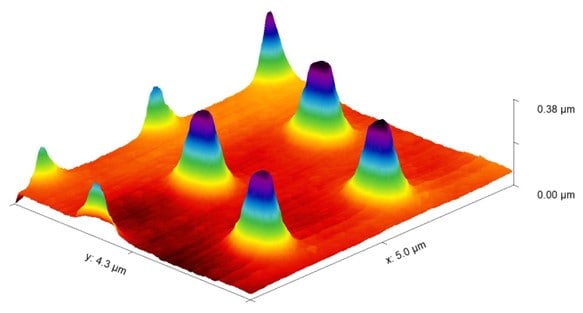
Figure 2: AFM image of pillars that are 500nm in diameter. These pillars were produced using UV lithography, they will be coated in a thin gold layer and used in Raman spectroscopy
These pillars once coated with a thin gold nanolayer can then be used for Raman spectroscopy to enhance the signal of biomarkers in the blood. In this application of Raman spectroscopy, a laser is directed towards the separated blood and light that is scattered off blood serum is then measured. The light that is scattered off the sample tells us which biomarkers are in present the sample, this is because the laser interacts with the molecules making them vibrate, wiggle and wag. Each molecule has a unique way of vibrating, and the concentration of these can then tell us about the presence and severity of a TBI at the earliest stages. The spontaneous Raman signal is very weak, and the aim of using these pillars is to improve the sensitivity of the diagnostic device.
A novel prognostic model of TBI has many possible applications. This approach to health monitoring could have many real-world impacts, including:
- Informing patients and family about expected outcomes
- Informing triage decisions, e., whether specialist care is required
- Improving design and analyses of clinical trials and trial data
- Benchmarking quality of care
- Improving monitoring of sub-acute and chronic conditions resulting from TBI
- Improving diagnosis of mild TBI at pitch-side, roadside, and bedside and monitoring long-term treatment
Figure 3: Optical microscope image of the ANMSA logo, University of Birmingham logo, and the lifETIME CDT logo
References
Rickard, J. J. S., Di-Pietro, V., Smith, D. J., Davies, D. J., Belli, A., & Oppenheimer, P. G. (2020). Rapid optofluidic detection of biomarkers for traumatic brain injury via surface-enhanced Raman spectroscopy. Nat Biomed Eng, 4(6), 610-623. https://doi.org/10.1038/s41551-019-0510-4