Revolutionizing Recovery: The Frontier of Free Flap Monitoring in Reconstructive Surgery
By LifETIME CDT Student: Narjes Meselmani (She/Her) (University of Galway)
This blog contains material of a highly sensitive nature including surgical procedures that may be triggering for some individuals.
Within the intricate realm of reconstructive surgery, specifically in the head and neck region, the success rate of free flap transplants serves as a remarkable monument to the progress of medical science. However, postoperative monitoring of these transplants is an essential aspect of this procedure. Traditionally, this monitoring has involved a combination of careful observation by medical professionals and technological assistance. However, recent innovative research is providing a novel aspect to this crucial stage: the utilization of machine learning-enhanced impedance monitoring.
Free flaps, which include the transfer of tissues from one area of the body to another, play a crucial role in reconstructive surgery, particularly following tumor removal, trauma, and some congenital defects with an aim to restore aesthetics, function, or both. The success of the procedure heavily relies on the viability of these flaps, which necessitates ensuring they receive sufficient blood supply. The existing methodologies, albeit efficient, possess certain constraints and usually catch ischemia too late, which puts the patient in distress and could lead to severe implications, including the need for more surgeries and sometimes death, hence creating opportunities for innovation and enhancement.
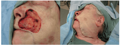
Trigger warning: Image of free flap reconstructive surgery
Here is where the innovative strategy of utilizing impedance monitoring in conjunction with machine learning algorithms becomes relevant. In this project, impedance probes, which assess the electrical characteristics of tissues, are currently being augmented with machine learning techniques to deliver more precise and up-to-the-minute information regarding the condition of free flaps. This approach not only guarantees to enhance the accuracy of postoperative monitoring but also creates opportunities for tailored patient care and proactive interventions.
The research involves thorough data collection and analysis. The deep learning models analyze impedance data obtained from biological tissues to identify patterns and features that may indicate potential difficulties, such as inadequate blood circulation or the onset of ischemia. This technique has the potential to completely transform the way surgeons and medical personnel assess the viability of free flaps, resulting in enhanced patient outcomes and a decreased likelihood of free flap failure.
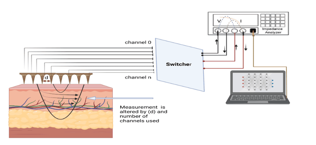
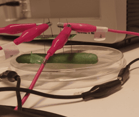
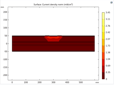
In the initial phase of this project, cucumbers were utilized as test subjects to confirm the concept’s validity. This involved observing the changes in their electrical properties, which could result from various factors such as dehydration. Additionally, computer simulations played a crucial role. They were employed to evaluate and refine the design of electrodes. This was done by analyzing the heat distribution and current density across different layers of the flap, ensuring optimal performance and accuracy in practical applications.
In conclusion, the goal of this project is to integrate machine learning with impedance probes for assessing the viability of free flaps following surgical procedures. This integration aims to improve postoperative care and patient outcomes. Leveraging the expanding reach of artificial intelligence, the project seeks to blend AI with medical devices to enhance healthcare results.